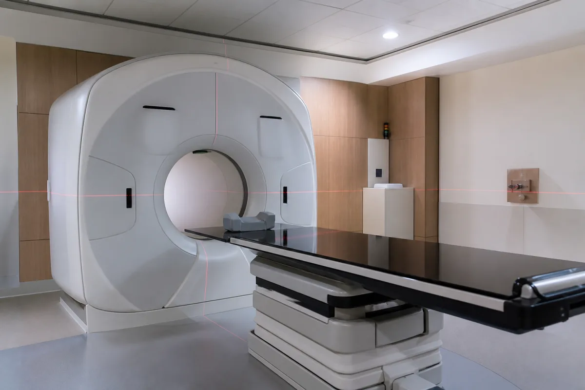
Radiation therapy CT simulator: a large-bore scanner with a flat couch top and external marking lasers, chosen by bore, scan field, tube power and laser accuracy
Large-bore CT simulators for radiotherapy planning scans · 82, 85 and 90 cm gantry openings against a 70 cm diagnostic bore, 50–70 cm scan fields, flat indexed couch top, movable wall lasers checked daily to ±2 mm
A radiation therapy CT simulator is a CT scanner set up for treatment preparation rather than diagnosis: the patient lies on a flat couch top in the immobilisation devices that will be used at the linac, the scanner acquires the planning images, and external lasers project reference lines onto the skin or the mask so the treatment isocentre can be marked. Three choices decide whether it works for a department: how wide the bore and the reconstructed scan field are, how much tube power and heat capacity it has for long thin-slice scans, and how accurately its lasers and couch top reproduce the patient's position.
CT simulators for external-beam radiotherapy: large-bore multislice CT scanners with a flat radiotherapy couch top, indexing for immobilisation devices, fixed or movable external positioning lasers and virtual simulation software, used to acquire planning CT images and mark the patient in the treatment position. This page is about the imaging and marking equipment in the simulation room. The planning software, dose-calculation engines and planning workstations that use these images are covered on the Treatment planning stations (radiotherapy TPS) page, and linac systems that plan and re-plan during the treatment session on the Integrated planning-and-delivery system for online adaptive radiotherapy page. Treatment couches at the linac, including robotic correction and the tabletop sag and travel between CT and treatment isocentres, are covered on the Six-degree-of-freedom radiotherapy treatment couch and Radiotherapy patient positioning couch pages; here the couch top appears only as part of the simulation room. Conventional fluoroscopic or radiographic simulators that mimic a linac gantry are described here only for their regulatory definition; diagnostic CT scanners without radiotherapy accessories are outside this page.
CNFX editorial research · Sources checked October 13, 2026 · 5 sources
Gantry opening of 82, 85 or 90 cm and the scan field that is actually reconstructed
The IAEA cites breast, lung and vulva patients scanned with their limbs held away from the torso, and head and neck patients in immobilisation devices, as cases where a 70 cm bore forces a change of position, giving a less reproducible set-up or a mismatch between imaging and treatment positions. [2] The IAEA gives 70 cm as the bore of conventional CT scanners (typ.), allowing objects up to 50 cm across (typ.), and describes large-bore scanners above 70 cm as designed for radiotherapy needs. [2] The 510(k) summaries below show that a wider gantry does not always come with an equally wide reconstructed field.
Canon's predicate in the same filing, a diagnostic Aquilion Prime SP, has a 780 mm opening, a 500 mm maximum field and ±30° tilt, which puts the 900 mm, no-tilt large-bore model in context. [5] Philips files its Big Bore in an Oncology configuration and a Radiology (Base) configuration, with intended use that includes images for oncology treatment preparation and radiation therapy planning. [3]
The IAEA warns that on some scanners CT numbers towards the periphery of large-field images are not reliable, which matters to dose calculation algorithms that convert CT numbers to density; it asks that the effect on dose calculation be evaluated at commissioning. [2] Editorial calculation: an 850 mm bore is 150 mm wider than a 70 cm bore, 75 mm more clearance on each side of the isocentre; the 85 cm Siemens models reconstruct 60 cm, leaving 25 cm of the opening outside the standard field.
Ask for the gantry opening and the maximum scan field with and without extended-field reconstruction, and for CT-number accuracy measured at the edge of the largest field you will use for planning.
02
Tube power and heat capacity for long planning scans at 3 mm slices
Planning scans cover longer volumes than diagnostic scans, preferably with 3 mm slices, and should be acquired quickly to limit motion; the IAEA names the heat storage capacity of single-slice CT tubes as the limit, lists the compromises otherwise made (fewer images, thicker slices, lower mAs or a higher pitch) and says greater heat storage capacity reduces them. [2]
Siemens lists maximum image acquisition lengths of 1600 mm and 2000 mm, depending on the patient table, for its go. platform scanners. [4] Editorial calculation: at the 3 mm slice thickness the IAEA prefers, a 2000 mm acquisition is about 667 contiguous images; Philips lists helical slice thicknesses from 0.67 to 5 mm and a 0.42 s rotation for its Big Bore. [3]
Give the supplier your longest planning scan (length, slice thickness, kV and mAs, 4D or not) and ask whether it runs in one acquisition without tube-cooling pauses.
03
Movable sagittal laser, indexed flat couch top and the ±2 mm daily checks
The IAEA treats a laser marking system as necessary for a CT used in radiotherapy, prefers at least the overhead sagittal laser to be movable, but puts alignment accuracy and stability ahead of movability, and asks for a laser line at least 50 cm long at the tomographic plane. [2] Fixed and movable wall lasers, it says, give the same quality of treatment. [2] Siemens includes control of RTP laser systems among the functions of its tablet workflow software. [4]
The in-gantry alignment lights of a diagnostic scanner are held only to ±5 mm, which the IAEA calls outside acceptable limits for radiotherapy; if they are used for marking they need to be tightened to ±2 mm, and on some scanners that is not sustainable because the lights sit on a vibrating fibreglass cover. [2] Couch checks are run with 70 kg and then 140 kg on the top to represent a patient. [2]
For the couch top itself, the IAEA asks that the CT and the linac tops share the same width, flex and sag under load and accept the same indexing of immobilisation devices, so the patient is set up in the same place on both. [2]
Ask which lasers are supplied (gantry, wall, ceiling; fixed or movable), their stated line width and positioning accuracy, and the couch top width, sag under 70 and 140 kg, and indexing pitch, compared with your linac couch.
04
Fluoroscopic simulator or CT simulator: two FDA device classifications
US regulation still has a separate class II device type, the radiation therapy simulation system, defined as a fluoroscopic or radiographic x-ray system used to localise the volume to be treated and confirm the position and size of the treatment field; it may include display equipment, patient supports and planning programs. [1] The large-bore scanners in this page's tables are instead cleared as computed tomography x-ray systems under 21 CFR 892.1750, product code JAK. [3][4]
Virtual simulation software takes over the conventional simulator's role on screen: the IAEA expects it to show gantry, couch, collimator and jaw motion, source–surface distance and beam divergence, to build digitally reconstructed radiographs, and to design portals with blocks and multileaf collimators. [2]
05
Data for a radiotherapy CT simulator
Paste it into your enquiry below or into an email to a maker, then replace every bracket.
Patients: [sites treated; widest set-up, e.g. breast board, arms up, head and neck mask]
Bore: [gantry opening; scan field, standard and extended]
Tube: [generator kW; heat capacity; longest planning scan and slice thickness]
Couch top: [flat top width; indexing; sag at 70 and 140 kg; linac couch to match]
Lasers: [gantry and wall lasers; movable sagittal laser; accuracy; control from the console]
Software: [virtual simulation; 4D or respiratory gating; DICOM export to the TPS]
Acceptance: [laser and couch checks; CT number to density at the largest field]
Explore company information and manufacturing capabilities, then discuss the scope of your project with a relevant supplier.
Share this page
Compiled and source-reviewed by the CNFX editorial team.
Each manufacturer above links to the page on that company's own website where the product is listed, with the date we checked it. How we source and review
· Page last updated 13 October 2026
Buyer questions
What bore size do radiotherapy CT simulators have?
Cleared large-bore models list gantry openings of 82 cm, 85 cm, 850 mm and 900 mm, against 70 cm for conventional CT in the IAEA's description. [4][3][5][2]
Is the scan field as wide as the bore?
No. The 85 cm Siemens go.Sim and go.Open Pro reconstruct 60 cm, the Philips 850 mm Big Bore up to 600 mm, and the 900 mm Canon Aquilion Exceed LB up to 700 mm with an extended-field option. [4][3][5]
How accurate must the marking lasers be?
The IAEA gives ±2 mm acceptable and ±1 mm achievable for external CT positioning lasers, checked daily by the radiographer and monthly by the physicist. [2]
Is a CT simulator the same device as a radiation therapy simulation system in US rules?
No. 21 CFR 892.5840 defines the simulation system as a fluoroscopic or radiographic x-ray system; large-bore CT simulators are cleared as CT x-ray systems under 892.1750. [1][3][4]
Sources and what still needs a direct check
Sources checked 2026-10-13. The US regulation that defines a radiation therapy simulation system, the IAEA's quality assurance programme for CT used in diagnosis and radiotherapy, and three FDA 510(k) summaries that list bore, scan field, tube and detector data for large-bore scanners.